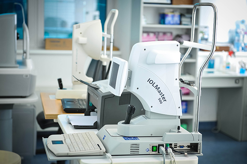
IOL master - A device that measures the strength of the lens to be fitted inside the eye during cataract surgery. Taking into account the length of the eye, ceratometric values, depth of anterior chamber and several other formulas it calculates the strength of the lens to be installed.
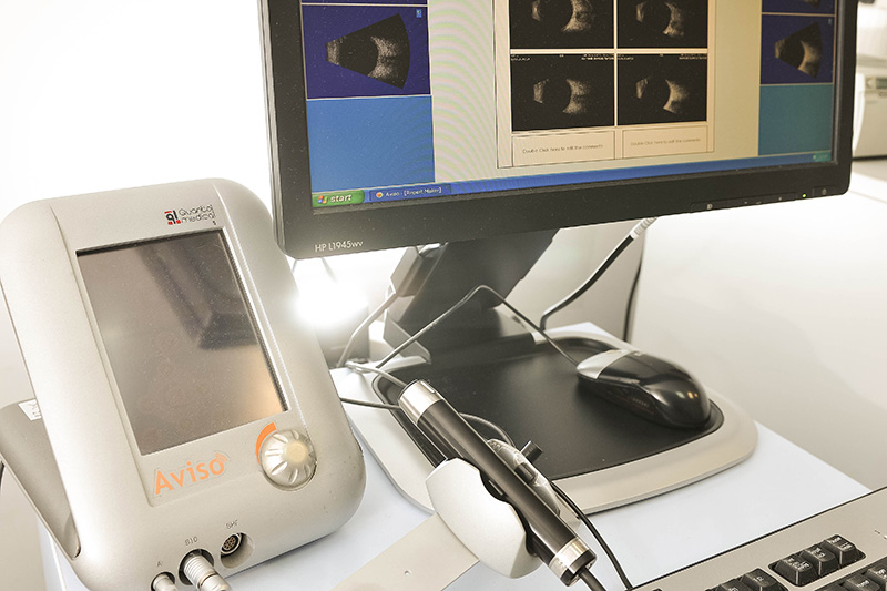
 Ultrasound + A scan
 - diagnostic procedure for obtaining access to the interior of the eye and the length of the eye.
How is it performed - the patient is sitting comfortably on a chair and the doctor with an ultrasound probe gently goes over the upper and the lower eyelids and takes photographs. When it comes to the A scan it is necessary to place the probe on the cornea of the previously anesthetized eye. This diagnostic procedure is painless, not harmful and it lasts only a few minutes.
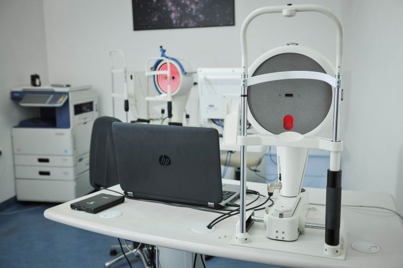
 Corneal topography
 - A test that gives an insight into the curvature of the front surface of the cornea and determines the corneal astigmatism.
How to do it - The patient sits on the device, lays his head and the device automatically records all the necessary data. This procedure is painless, lasts only a few seconds, and the result is obtained immediately and printed out on the printer.
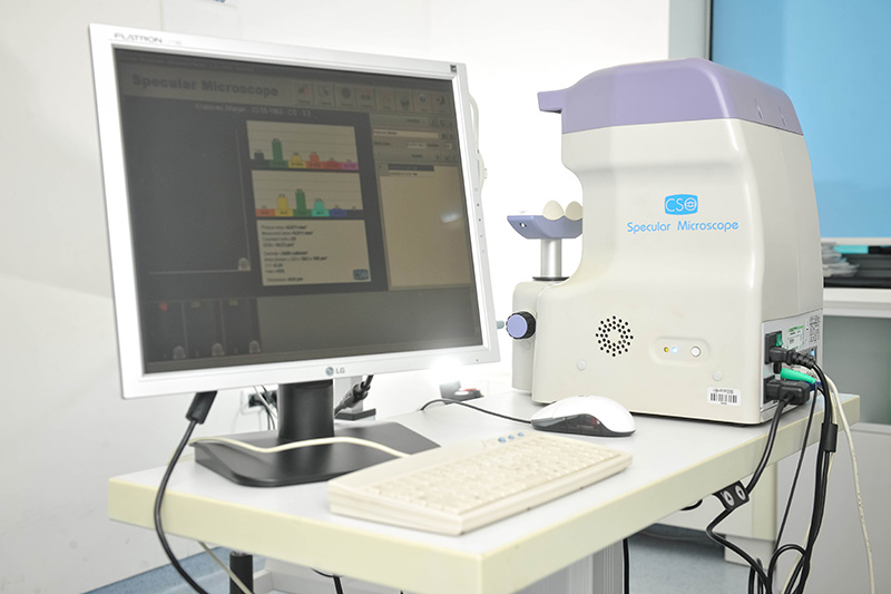
 Endothelial Microscope
 - A test that calculates the number of endothelial cells in the cornea.
How to do it - The patient sits on the device, lays his head and the device automatically records all the necessary data. This procedure is painless, lasts only a few seconds, and the result is obtained immediately and printed out on the printer.
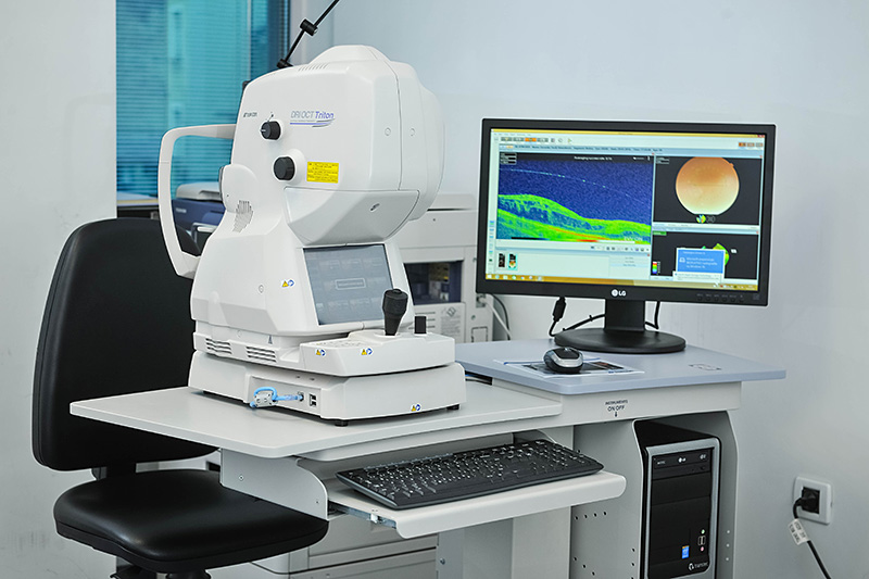
 Optic coherence tomography of macula and optic nerve
 - Captures the structure of macula and optic nerve and reveals possible abnormalities in these structures, otherwise invisible code review with a magnifying glass.
How to do it? The patient sits on the device, places his head on it and the device automatically records all the necessary data. This procedure is painless, lasts only a few seconds, and the result is obtained immediately and printed out on the printer.

 Ultrasound + A scan - diagnostic procedure for obtaining access to the interior of the eye and the length of the eye.
 Corneal topography - A test that gives an insight into the curvature of the front surface of the cornea and determines the corneal astigmatism.
 Endothelial Microscope - A test that calculates the number of endothelial cells in the cornea.
 Optic coherence tomography of macula and optic nerve - Captures the structure of macula and optic nerve and reveals possible abnormalities in these structures, otherwise invisible code review with a magnifying glass.