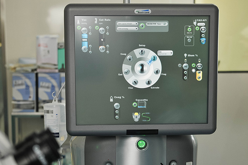
Stellaris Bausch and Lomb
The platform for a minimally invasive vitrectomy using 23 and 25 gauge incisions, high "cut rate" 5000 cpm, combined with the unit for cataract surgery and two sources of light.

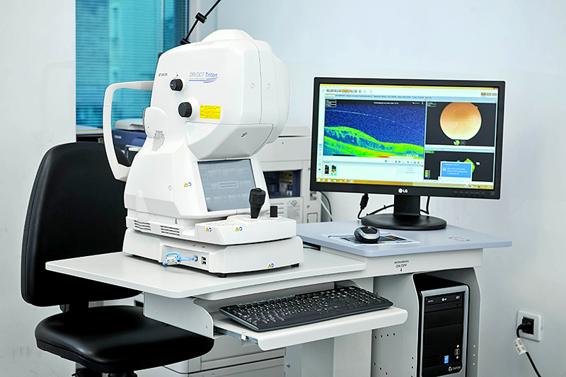
Optical coherence tomography OCT - Topcon Swept Source
Optical coherence tomography for the analysis of the macula and retina with 100,000 scans per second and the penetrability of scans for the analysis of the retina and choroid. Svjetlost is among few to posses the highest resolution OCT device in the world, the only in this part of Europe. Now with a new software update we can perform OCT angiography to scan eye vasculatore without dye injection.

How is the test performed?
A patient places his or her head on a chin rest and the device automatically captures the fundus image. The test is painless, only few seconds in duraiotn and is completely harmlesssince the patient is not exposed to any radiation. The image is printed immediately. The test can also be performed with a narrow pupil.

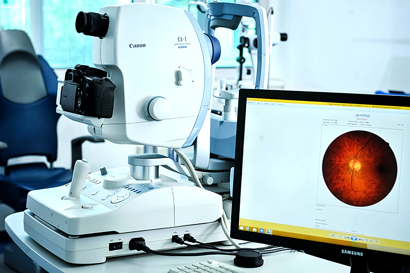
Fluorescein angiography Canon 12 megapixel camera
Fluorescein angiography is a test based on the analysis of the eye blood vessels and is used in diagnosis of macular degeneration, diabetic retinopathy and some other diseases of the retina. The test is performed less often after the introduction of OCT, but it can still be crucial in establishing the diagnosis.
How is the test performed?
A dye is injected intavenously and the retina of both eyes is photographed for a few minutes. .Discomfort during the test is minimal and is due to injection of dye into the vein. Yellowish discoloration of the skin is possible and goes away within few. The images are printed immediately ad can be analysed right away. Our camera has a high resolution of 12 mega pixels and allows taking images of the retina in the autofluorescence mode as well.
Echography of the eye -Quantel Medical
Ultrasound of the eye is a valuable test that gives an insight into the interior of the eye in the event that optical media are not transparent, for example with cataracts or bleeding. It also supplements the clinical findings and confirms the diagnosis in a number of retinal diseases. It is most oftenly used in the diagnosis of the bleeding in the eye, eye injuries, suspicion of foreign eye bodies, retinal detachment, ocular and orbital tumours.
How is the test performed?
A patient is comfortably placed in a chair. A probe is gently pressed against eye lids to scan from different angles. It is painless, harmless and of few minutes duration.
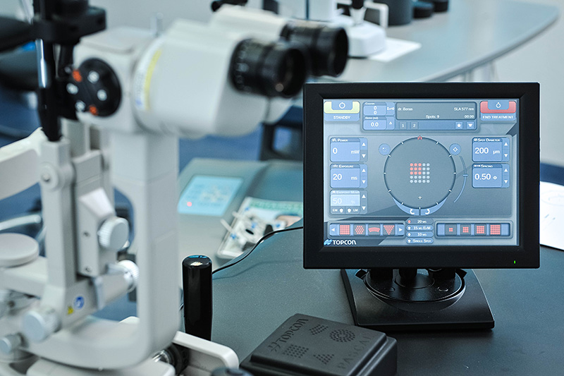
Topcon Pascal laser photocoagulation
This is a new generation laser for the treatment of retinal diseases. Unlike the previous lasers, the treatment is significantly less painful and of a shorter duration. The laser fires high number of laser spots in a shorter interval and the retinal burn is of a lower intensity.


